Radiographic positioning terminology

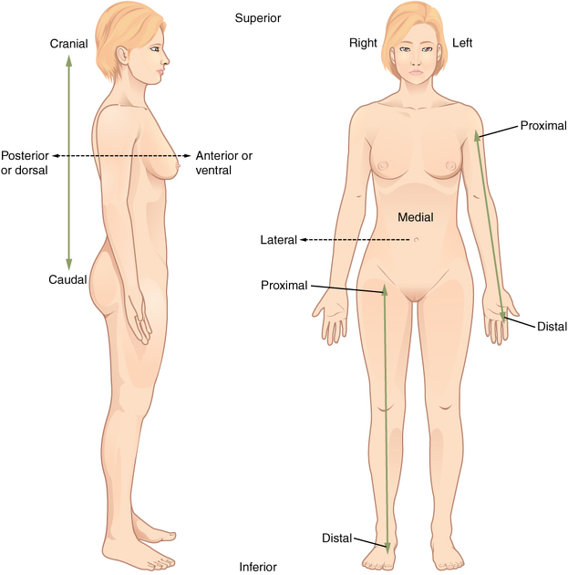
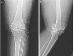
This is used to describe the position of the patient for taking various radiographs. Standard nomenclature is employed with respect to the anatomic position.

Basic terms of relations
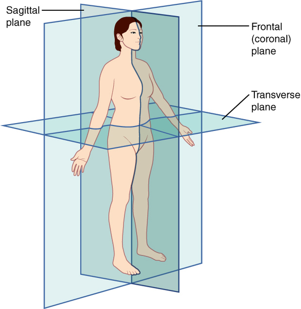
Planes
  • the axial plane (transverse or transaxial plane): horizontal plane perpendicular to the long axis of the body
    • divides the body into superior and inferior parts
  • the sagittal plane: vertical plane parallel to the median plane (or midsagittal plane)
    • divides the body into right half and left halves
  • the coronal plane: vertical plane perpendicular to the median plane
    • divides the body into anterior and posterior parts
BODY POSITION
  • Erect: either standing or sitting
  • Decubitus: lying down
  • Supine: lying on back
  • Trendelenburg position: the patient is supine (on an inclined radiographic table) with the head lower than the feet
  • Prone: lying face-down
  • Lateral decubitus: lying on one side
    • right lateral: right side touches the cassette
    • left lateral: left side touches the cassette
Movement
  • flexion: decrease in the angle of the joint
  • extension: increase in the angle of the joint
  • abduction: movement of limb away from midline
  • adduction: movement of limb towards the midline
  • pronation: movement of hand and forearm to bring the palm facing posterior
  • supination: movement of hand and forearm to bring the palm facing anterior
  • circumduction: circular movement of a joint using a combination of flexion, abduction, extension and adduction such that the distal limb describes a circle
  • opposition: thumb brought to oppose another digit
  • reposition: thumb repositioned back to the anatomic position
  • elevation: movement of the scapular superiorly
  • depression: movement of the scapular inferiorly
  • eversion: movement of the sole of the foot away from the median plane
  • inversion: movement of the sole of the foot towards from the median plane
  • protrusion: movement of the mandible, lips or tongue anteriorly
  • retraction: movement of the mandible, lips or tongue posteriorly

Projections

  • antero-posterior (AP)central ray passes, perpendicular to the coronal plane, from anterior to posterior
  • postero-anterior (PA): central ray passes, perpendicular to the coronal plane, from posterior to anterior
    • depending on the anatomic segment to radiograph, synonyms can be used, for example: occipito-frontal (skull); dorso-ventral (thorax); dorso-palmar (hand)
  • lateral: central ray, perpendicular to the sagittal plane and parallel to the coronal plane, passes from one side of body to the other
  • oblique: central ray passes through the body/body part through a plane which is at an angle to the transverse plane/coronal plane
  • axial: central ray passes through (or parallel) to the long axis of the body
    • in some cases, however, the central ray runs through (or parallel) to the long axis of the skeletal segment studied (for example, the axial view of the calcaneus)

Welcome to Imaging Technology Gallery

X-RAY MACHINE
Computed Tomography machine
X-ray machine
PROCESS OF RECORDING IMAGES
X-RAY TUBE
medical-imaging-equipment-and-accessories

Depressed?

depression mental health brain dra martha castro noriega mexico tijuana
There are solutions to the tricky depression. Keep reading, you will find more useful tips.

Try as much as possible to avoid been depressed. Though it is inevitable, but can be managed properly by yourself only. How?

What Does Depression Really Feel Like?

Depression symptoms vary from person to person, as do the severity of the symptoms. However, C=common symptoms associated with depression include:

Feeling Sad, Empty, Or Anxious. While everyone experiences feelings of sadness from time to time, it’s important to understand that depression goes beyond feeling sad. People living with depression may face these symptoms regularly and for long periods of time with little or no improvement.

Irritability. Feeling restless, being more easily agitated, or feeling cranky may all be responses to the helplessness that often accompanies depression.

Loss Of Interest. A person living with depression may be disinterested in hobbies or activities that used to bring enjoyment. A decrease in or absence of desire for sexual intimacy may also be noticeable and can be a sign of depression.

Difficulty Concentrating. Simple things like reading a magazine or watching a TV series may become boring to a person experiencing depression. Or, you may experience an inability to focus on a task. It may also feel overwhelming to make decisions. 

Changes In Appetite. Some people who experience depression may eat, even if they aren’t hungry. Others experience loss of appetite entirely and, in some cases, may skip several meals without thinking about it. 

Alterations In Sleep Patterns. While people experiencing depression may wake up too early or have trouble falling asleep, the opposite can also happen, resulting in sleeping much longer than usual.

Depression can be a tricky beast. It usually creeps in slowly, so by the time you’ve realized it’s upon you, you feel like it’s about to consume you. Anyone who has experienced depression in the past or who goes through bouts of depression currently will tell you how tough it is to navigate symptoms.

“Talking to a mental health professional offers you an opportunity to talk with someone who can guide you on ways to improve and manage your depression symptoms.”

Sometimes it can be difficult to determine why you are feeling depressed. The cause of depression is varied, as is the cure. If you’re wondering, “Why am I always depressed?” you’re not alone. We’ve developed a list of possible depression causes, along with suggestions about strategies you may consider using to reclaim your life from deep depression and start feeling like yourself again.

Causes Of Depression

There is no single cause for depression and a depression diagnosis depends on a combination of dynamics such as a person’s environmental conditions, as well as their genetic make-up. That said, doctors consider several factors to determine a diagnosis of depression. A few examples of these factors include:

  • History of depression in the family
  • History of other mental health diagnoses (such as anxiety, PTSD, or bipolar disorder)
  • Hormone changes (such as menstrual cycles and pregnancy)
  • History of experiencing a traumatic event (such as abuse or violence)
  • The use of medications (such as sleeping aids or blood pressure medication)
  • Substance use or abuse, past or present

Overcoming Depression

As stated above, depression is not just a temporary feeling of sadness. It is a disorder that takes time to treat and manage. But it’s encouraging to know that it is possible to overcome depression symptoms and live a happy life. Non-medical means of overcoming depression symptoms include being proactive with self-care efforts, such as:

  1. Bathing or showering daily
  2. Getting dressed (even if you don’t feel like it)
  3. Eating a well-balanced diet
  4. Exercising regularly to the point of sweating

Although these may seem like somewhat menial tasks, completing them each day can help improve your overall mood and feelings. 

Consult With Your Doctor

While there are many helpful resources to collect information about depression, such as those found in this article, an accurate diagnosis should only be made by a physician or mental health professional. If you are experiencing depression symptoms, consider making an appointment with your primary care provider for an initial evaluation.

Depression Can Cause Permanent Brain Damage!
Depression not only makes a person feel sad and dejected – it can also damage the brain permanently, so the person has difficulties remembering and concentrating once the disease is over. Up to 20 percent of depression patients never make a full recovery.

PROPOSAL SOLUTIONS:

The mere fixation on anti-depressants will not solve this problem as the inflammation is very probably not limited to the brain. Silent inflammation is becoming more and more visible as the driving force behind chronic diseases.

Solutions:
An anti-inflammatory lifestyle with antioxidant-rich nutrition, physical activity such as exercise and avoidance of toxins are the logical conclusion for starters. Always engage in physical fitness exercise, this also helps a lot. Thanks for visiting!

https://pagead2.googlesyndication.com/pagead/js/adsbygoogle.js

X-Ray Tube

The x-ray tube serves the function of creating x-ray photons from electric energy supplied by the x-ray generator. The process of creating the x-ray beam is very inefficient, with only 1% of the electric energy converted to x-ray photons and the remaining 99% converted to heat in the x-ray tube assembly. Thus, to produce sufficient x-ray output for diagnostic imaging, the x-ray tube must withstand and dissipate a substantial heat load, a requirement that affects the design and composition of the x-ray tube. The major x-ray tube components are the cathode and anode assemblies, the tube envelope, the rotor and stator (for rotating anode systems), and the tube housing. The design of the x-ray tube determines the basic characteristics of the x-ray beam such as focal spot size, x-ray field uniformity, and the x-ray energy spectrum. These x-ray beam characteristics are important because they affect radiologic parameters such as spatial resolution, image contrast, and patient dose.

What is an abdominal CT scan? Definition. Uses. What to expect.

Computed tomography (CT) is a type of medical imaging tool that allows doctors to see inside a person’s body.Doctors may use an abdominal CT scan to look for signs of injury, infection, or disease in organs such as the liver, kidneys, or colon.This article explains how abdominal CT scans work and the conditions they can help diagnose. It also outlines what preparations to make before having a CT scan and what to expect during the procedure.

The CT scanner uses a thin beam of radiation to scan the body from many different angles. It then feeds the scanned images into a computer, which creates individual slices, or cross-sections, of the inside of the body. The American Cancer Societylikens these cross-section images to the individual slices of a loaf of bread.

A CT scanner can also layer all the scanned images on top of one another to create a 3D image of the scanned body area. Specialist doctors called radiologists can then rotate the 3D image on a computer screen, to view it from different angles.

Uses

Doctors may use a CT scan to help diagnose the cause of pain in the abdomen or pelvic area. Some conditions that may cause pain in these areas include:

Other potential uses for a CT scan include:

  • helping to identify any internal bleeding or injuries following an accident
  • planning surgery, and checking the results of the operation
  • helping to guide a needle during a needle biopsy
  • helping a doctor to plan and administer radiation therapy
  • helping a doctor to check thatchemotherapy is working

What to expect

Below is some information on what people can expect before and during their abdominal CT scan.

Before the scan

Below are some procedures that people may need to follow before they go in for their CT scan.

Fasting

When diagnosing bowel conditions, a doctor will need a clear view of the inside of a person’s bowel. As such, they may tell a person to avoid eating or drinking for several hours before their scan. Alternatively, a person may receive a laxative or enema to clear out the bowel.

Taking a contrast dye

In some cases, doctors will ask a person to take a contrast dye before their CT scan. The dye helps the internal organs and blood vessels show up more clearly on the scan. A person may take the dye orally or receive it through a vein or an enema.

The contrast dye is not suitable for everyone. People should tell their healthcare team if they are pregnant or have ever experienced an allergic reaction to any of the following:

Removing metal objects

Before having a CT scan, a person will need to undress and put on a gown. They will also need to remove any metal objects, as these can interfere with the CT images. Such objects may include:

  • jewelry
  • hairpins
  • spectacles
  • dentures
  • underwired bras
  • belts with metal belt buckles

Anyone who has an implanted medical device, such as a pacemaker or infusion port, should tell their healthcare team about the device before the scan.

During the scan

A CT scanner is a large, donut-shaped machine with a tunnel in the middle where the scanning takes place. A person lies on a flat table that slides in and out of the tunnel. Sometimes, the medical team may use pillows or straps to keep the person in the correct position, while the scan is taking place. 

The person will need to stay perfectly still for the duration of the scan, which usually only takes a few minutes. It does not hurt, but some people may find it uncomfortable to stay still for this amount of time.

Risks

Some people have concerns about the potential risks of CT scans. We outline two key concerns below.

Increased cancer risk

Some people worry that CT scans will increase their risk of developing cancer. This is because CT scanners use a form of ionizing radiation to image the inside of the body.

It is worth remembering that a CT scan only delivers a low dose of radiation, and the cancer risk is small. According to the National Cancer Institute, not having the procedure can be much riskier than having it. This is especially so if a doctor is using the CT scan to help diagnose cancer or another serious condition.

Contrast dye allergies

Some people may experience a reaction to the contrast dye they take before the CT scan. The side effects are usually mild. However, they can be severe in rare cases.

Potential mild side effects include:

Potential severe side effects include:

Anyone who experiences any severe side effects should tell their healthcare team straight away.

To help avoid the risk of side effects, people should tell their doctor if they:

SUMMARY

A CT scan is a medical imaging tool that allows doctors to see bone, organs, and soft tissues inside a person’s body. Doctors may use an abdominal CT scan to look for signs of injury, infection, or disease in organs such as the colon, spleen, liver, or kidneys.

A CT scan usually takes only a few minutes. The procedure does not hurt, but some people may find it uncomfortable to lie still for the duration of the scan. A doctor will usually receive the results of a CT scan within a few days. People will need to make a follow-up appointment with their doctor to discuss the results of the scan. They can also take this opportunity to talk through their diagnosis and any treatments they may need

Knowledge is power. Get our free daily publication on radiography and healthcare by visiting our site regularly. Thank you for visiting.

MRI Scan

Contents

  1. Overview
  2. How it’s performed
  3. Who can have one
Picture of a person having an MRI scan

Magnetic resonance imaging (MRI) is a type of scan that uses strong magnetic fields and radio waves to produce detailed images of the inside of the body.

An MRI scanner is a large tube that contains powerful magnets. You lie inside the tube during the scan.

An MRI scan can be used to examine almost any part of the body, including the:

  • brain and spinal cord 
  • bones and joints 
  • breasts 
  • heart and blood vessels 
  • internal organs, such as the liver, womb or prostate gland  

The results of an MRI scan can be used to help diagnose conditions, plan treatments and assess how effective previous treatment has been.

What happens during an MRI scan?

During an MRI scan, you lie on a flat bed that’s moved into the scanner. 

Depending on the part of your body being scanned, you’ll be moved into the scanner either head first or feet first.Credit: 

The MRI scanner is operated by a radiographer, who is trained in carrying out imaging investigations. 

They control the scanner using a computer, which is in a different room, to keep it away from the magnetic field generated by the scanner.

You’ll be able to talk to the radiographer through an intercom and they’ll be able to see you on a television monitor throughout the scan.

At certain times during the scan, the scanner will make loud tapping noises. This is the electric current in the scanner coils being turned on and off. 

You’ll be given earplugs or headphones to wear.

It’s very important to keep as still as possible during your MRI scan. 

The scan lasts 15 to 90 minutes, depending on the size of the area being scanned and how many images are taken.

How does an MRI scan work?

Most of the human body is made up of water molecules, which consist of hydrogen and oxygen atoms. 

At the centre of each hydrogen atom is an even smaller particle called a proton. Protons are like tiny magnets and are very sensitive to magnetic fields.

When you lie under the powerful scanner magnets, the protons in your body line up in the same direction, in the same way that a magnet can pull the needle of a compass.

Short bursts of radio waves are then sent to certain areas of the body, knocking the protons out of alignment. 

When the radio waves are turned off, the protons realign. This sends out radio signals, which are picked up by receivers.

These signals provide information about the exact location of the protons in the body. 

They also help to distinguish between the various types of tissue in the body, because the protons in different types of tissue realign at different speeds and produce distinct signals. 

In the same way that millions of pixels on a computer screen can create complex pictures, the signals from the millions of protons in the body are combined to create a detailed image of the inside of the body.

Safety

An MRI scan is a painless and safe procedure. You may find it uncomfortable if you have claustrophobia, but most people are able to manage it with support from the radiographer. 

Going into the scanner feet first may be easier, although this isn’t always possible.

Extensive research has been carried out into whether the magnetic fields and radio waves used during MRI scans could pose a risk to the human body. 

No evidence has been found to suggest there’s a risk, which means MRI scans are one of the safest medical procedures available.

But MRI scans may not be recommended in certain situations. For example, if you have a metal implant fitted, such as a pacemaker or artificial joint, you may not be able to have an MRI scan. 

They’re also not usually recommended during pregnancy.